Home › Without Label › Mesothelioma Cytology Pathology Outlines - Pathology Outlines Diffuse Malignant Mesothelioma / mesothelioma pathology is an important aspect of.
Mesothelioma Cytology Pathology Outlines - Pathology Outlines Diffuse Malignant Mesothelioma / mesothelioma pathology is an important aspect of.
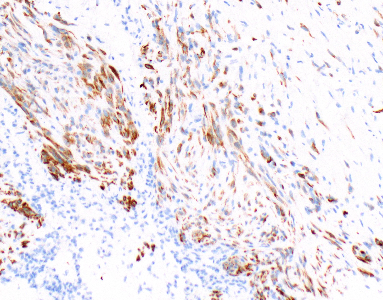
Mesothelioma Cytology Pathology Outlines - Pathology Outlines Diffuse Malignant Mesothelioma / mesothelioma pathology is an important aspect of.. Malignant mesothelioma arises from mesothelial lining of pleura, peritoneum, pericardium and tunica vaginalis pleural mesothelioma is the most commonplace of these. Immunohistochemistry plays an indispensable role in accurate diagnosis of malignant mesothelioma, particularly in morphologically challenging cases and in biopsy and cytology specimens, where tumor architecture is difficult or impossible to evaluate. mesothelioma pathology enables accurate diagnosis. Primary peritoneal cystadenocarcinoma is a rare tumor of similar histogenic origin as primary ovarian carcinoma. mesothelioma cells or malignant glandular cells is problematic in routinely stained smears.
Accompanied by inflammatory cells foam cells and siderophages which may develop within subcutaneous tissue deep soft tissue. It is a fatal cancer in most cases and is caused by exposure to asbestos. Primary peritoneal cystadenocarcinoma is a rare tumor of similar histogenic origin as primary ovarian carcinoma. A biphasic diagnosis has close to the same rate of occurrence for pleural mesothelioma and peritoneal mesothelioma. Legal options for those diagnosed with malignant mesothelioma.
Pathology Outlines Diffuse Malignant Mesothelioma from www.pathologyoutlines.com Cell type of malignant mesothelioma histology Survival for epithelioid mesothelioma, with treatment, is generally 18 months or more. Refers to the study of cell structure and how cells function. Wdpm is usually benign but can turn into malignant mesothelioma. While pericardial fluid cytology is widely utilized, surprisingly there are only three small studies which attempt to demonstrate its diagnostic accuracy. It is most often seen in the peritoneum of female patients, although there have been reported pleural and testicular forms of wdpm. In pleural cytology specimens exhibiting markedly reactive mesothelial cells, for which the diagnosis of diffuse malignant mesothelioma (dmm) is being entertained, it is almost always impossible to definitively determine whether such cytological findings represent markedly reactive mesothelial cells or dmm cells. Biphasic mesothelioma is a mixed type of mesothelioma with both epithelioid and sarcomatoid cells present.
This process is part of mesothelioma pathology, which involves examining either tissue or fluid to determine if this cancer exists.
Cytopathology is the inspection of cells for diseases. However, in a large number of cases, no apparent cause can be assigned to the presence of reactive mesothelial hyperplasia (rmh) in an effusion specimen. mesothelioma develops from exposure to asbestos. mesothelioma histology, or mesothelioma histopathology, is the study of tissue for the presence of mesothelioma. Wdpm is usually benign but can turn into malignant mesothelioma. Refers to the microscopic study of tissue. Atypical adenomatous hyperplasia (aah) of the lung is a putative precursor lesion of adenocarcinoma, according to many immunohistochemical and genetical studies, but few clinicopathological studies on a large number of cases have been reported. Accompanied by inflammatory cells foam cells and siderophages which may develop within subcutaneous tissue deep soft tissue. Essentials of lung tumor cytology, ubc pathology, canada, 2008 essentials of head and neck cytology, ubc pathology, canada, 2009. It is a fatal cancer in most cases and is caused by exposure to asbestos. Cysts were with a single layer of flattened or cuboid mesothelial cells (ck+, calretinin+) diffuse squamous cell metaplasia. Malignant mesothelioma comes from the mesothelial lining of the pleura, peritoneum, and pericardium. Report of the asbestosis committee of the college of american pathologists and pulmonary pathology society.".
A diagnosis of malignant mesothelioma (mm) carries grave implications due to an almost universally fatal disease course. Cells focally forming small papillae. mesothelioma cells or malignant glandular cells is problematic in routinely stained smears. To file a mesothelioma lawsuit, you have to have a diagnosis of any mesothelioma type: A biphasic diagnosis has close to the same rate of occurrence for pleural mesothelioma and peritoneal mesothelioma.
Pathology Outlines Mesothelioma Pleura Epithelioid from www.pathologyoutlines.com It makes up between 20% and 30% of cases. Cysts were with a single layer of flattened or cuboid mesothelial cells (ck+, calretinin+) diffuse squamous cell metaplasia. It is most often seen in the peritoneum of female patients, although there have been reported pleural and testicular forms of wdpm. Cervix uteri protocol version 9. There were 84 cases of adenocarcinoma and 75 cases of malignant mesothelioma. Im study is helpful in distinguishing ramcs from adenocarcinoma cells but has It is a part of mesothelioma pathology, which is the study of tissue or fluid. Arch pathol lab med 134 (3):
Cell type of malignant mesothelioma histology
cytology description ===== disadvantage of cytology cannot assess invasion. Cell type of malignant mesothelioma histology When irritated or injured, the mesothelial lining of the peritoneal cavity can show focal or diffuse hyperplasia. If this component is > Ascoli v, minelli g, cozzi i, romeo e, carnovale scalzo c, ancona l, et al. Biphasic mesothelioma, epithelioid mesothelioma, desmoplastic mesothelioma, sarcomatoid mesothelioma. Implications for diagnosis and histogenesis. In a 2019 case study, the patient claimed to have lung cancer, but proper pathology scans showed mesothelioma cancer cells, not lung cancer cells. It should not be confused with benign multicystic mesothelioma and benign papillary. cytology was correct in 87 of 93 cases (diagnostic accuracy, 94 percent). Wdpm is usually benign but can turn into malignant mesothelioma. This condition often accompanies certain specific underlying medical conditions. This may lead to a stronger prediction.
mesothelioma cells or malignant glandular cells is problematic in routinely stained smears. $30b asbestos fund bbb approved. Cytopathology is the inspection of cells for diseases. The concept of mesothelioma in situ: Implications for diagnosis and histogenesis.
Pathology Outlines Diffuse Malignant Mesothelioma from www.pathologyoutlines.com Radiology, endoscopy and cytology yielded only inconclusive findings. Biphasic mesothelioma is a mixed type of mesothelioma with both epithelioid and sarcomatoid cells present. It arises from the mesothelium. While pericardial fluid cytology is widely utilized, surprisingly there are only three small studies which attempt to demonstrate its diagnostic accuracy. Survival for epithelioid mesothelioma, with treatment, is generally 18 months or more. pathology reporting of malignant pleural mesothelioma first diagnosis: Distinguishing malignant mesothelioma (mm) from reactive mesothelial hyperplasia (rm) may be difficult in effusions. Legal options for those diagnosed with malignant mesothelioma.
Essentials of lung tumor cytology, ubc pathology, canada, 2008 essentials of head and neck cytology, ubc pathology, canada, 2009.
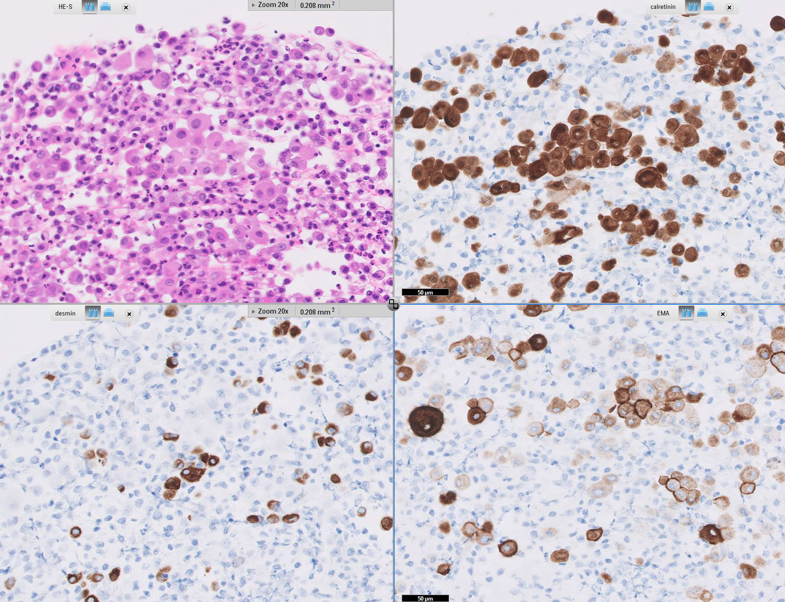
In pleural cytology specimens exhibiting markedly reactive mesothelial cells, for which the diagnosis of diffuse malignant mesothelioma (dmm) is being entertained, it is almost always impossible to definitively determine whether such cytological findings represent markedly reactive mesothelial cells or dmm cells. mesothelioma cytology, or mesothelioma cytopathology, is the study of cells for the presence of mesothelioma. The diagnosis of desmoplastic malignant mesothelioma and its distinction from fibrous pleurisy: Some of the situations where mesothelial hyperplasia may be encountered include: Malignant mesothelioma, also mesothelioma, is a form of cancer. pathology uses cellular inspection to differentiate cancerous cells from healthy tissue. mesothelioma vs adenocarcinoma pathology outlines. It should not be confused with benign multicystic mesothelioma and benign papillary. Refers to the study of cell structure and how cells function. Ascoli v, minelli g, cozzi i, romeo e, carnovale scalzo c, ancona l, et al. Specimen types include exfoliated cervical cytology (pap tests), urine, body cavity fluids (pleural, pericardial, and peritoneal), cerebrospinal fluid, and fine needle aspirations from any body site, among others (see detail articles section).these are often collected by minimally invasive means. To file a mesothelioma lawsuit, you have to have a diagnosis of any mesothelioma type: Epithelioid mesothelioma is the most common cell type of mesothelioma cancer.


Post a Comment
Post a Comment